
 Tiếng Việt


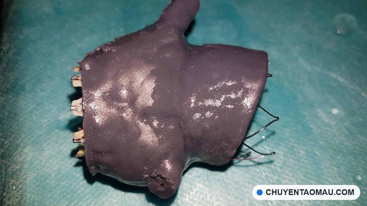
The first anatomical model I printed for a doctor was a section of a lower jaw, rebuilt from the patient’s CT images. There was nothing fancy about it: white resin, about six hours of print time. But when the doctor picked it up, turned it around and said, “Now I know where to cut,” I realized 3D printing in medicine has a very different kind of value from printing an enclosure or a display model.
This month brought quite a few notable stories on biomedical 3D printing, both in Vietnam and abroad. Put together, they give a fairly clear picture of what has been achieved and what still has to wait.

Artificial blood vessels: a Vietnamese team masters its own bioprinting system
According to VnExpress, the research team of Assoc. Prof. Dr. Nguyen Ngoc Dinh (University of Science, Vietnam National University, Hanoi) spent more than two years building and mastering a complete 3D bioprinting system, working with VinUni and the Vietnam Military Medical University. The team uses the Kenzan method: cell spheroids are skewered onto a “needle array,” which requires placing needles to an accuracy of 0.001 mm. The 3D-printed blood vessels were trial-grafted into the abdominal aorta of lab rats, and all the rats kept normal blood flow and stable blood pressure.
The context is what makes this research matter: according to the Ministry of Health, Vietnam has about 8,000–9,000 people who need a kidney transplant, 10,000 who need a liver transplant and 1,000 who need a heart transplant. Dr. Than Thi Trang Uyen of the team made a point I strongly agree with: if we only import machines, local teams will just be operators of a closed foreign ecosystem.
Still, Assoc. Prof. Do Xuan Hai was frank that regulatory hurdles could take 5–10 years. The earliest likely applications are ear cartilage, knee cartilage, skin and tissue for drug testing. In parallel, another team at Vietnam National University, Ho Chi Minh City has printed skin tissue using bioinks made from gelatin, alginate and collagen, aimed at treating burns and hard-to-heal wounds, and at drug and cosmetics testing.

Braided 3D-printed nitinol stents: they bend without breaking
In Europe, 3D Printing Industry reports that the IMDEA institute and the Technical University of Madrid (UPM), with MIT taking part, have 3D printed nitinol stents with a braided, woven-like structure. The stent can be compressed to 50% of its diameter and still recover more than 90% of its shape. The pressure on the vessel wall is up to 5 times lower than with conventional 3D-printed lattice stents, which risk breaking at around 25% strain.
For engineers, the nice thing here is how geometric design solves a material problem. Nitinol is superelastic, but a rigid lattice creates stress concentrations. Switching to a braided structure lets the “strands” slide slightly over each other and spread the strain. This is something only 3D printing can do easily. Even so, the team says the stent hasn’t been tested on living tissue yet; the next step is in vitro testing, then animal trials.
CE-certified dental resins: an application already on the market
If blood vessels and stents are still in the lab, dentistry is the field where medical 3D printing is most commercialized. In late September, AmeraLabs introduced three CE Class I dental resins: one for impression trays, one for implant surgical guides with a Shore D hardness of 85 that can be autoclaved, and one for try-in teeth with a flexural modulus above 2 GPa. They cure at 385/405 nm, work on many popular resin printers, and meet the ISO 10993-1 biocompatibility standard.


3D data: the foundation of every medical application
One story isn’t strictly about 3D printing, but I find it very relevant. VnExpress described a system that builds 3D models from CT images and overlays them on live X-ray images to guide aortic stent placement. Each scan takes only 5–20 seconds, and from 2023 to August 2026 the system supported more than 1,600 cases at a hospital in Hanoi.
What all of these have in common: everything starts with accurate 3D data. Whether you’re printing an anatomical model, a surgical guide or a bioprinted blood vessel, turning CT/MRI images into a clean, correctly sized 3D model is still what makes or breaks the result.

An engineer’s view: where should Vietnam start?
From my experience working with clinics and hospitals, I see three areas where 3D printing can contribute right now, without waiting 5–10 years:
- Anatomical models for surgical planning and for explaining procedures to patients.
- Surgical guides in dentistry and orthopedics, using materials that are already certified.
- Personalized assistive devices and splints made from 3D body scan data.
Bioprinting is a long-distance race, and a local research team mastering the core technology is a very encouraging sign. While the regulatory framework catches up, more “modest” applications can already help doctors operate more precisely and patients recover faster, starting today.


About CT3D Group and In3D.edu.vn
CT3D Group supplies resin 3D printers and high-precision 3D scanners, and offers anatomical model printing and rapid prototyping services for healthcare, dentistry and manufacturing. CT3D engineers support every step, from processing CT/3D scan data to printing and finishing.
Doctors and technicians who want to run the workflow themselves can check out the courses at 3D printing training – In3D.edu.vn. See more solutions at CT3D Group – 3D printing and 3D scanning technology.
Sources: VnExpress (Jun 30, Jun 4 and Oct 7, 2026), 3D Printing Industry (Oct 20 and Sep 29, 2026).
Compiled by Nguyen Cuong
Frequently asked questions
How is bioprinting different from printing a regular anatomical model?
Bioprinting uses bioinks containing living cells or biocompatible materials to create tissue that can survive and integrate into the body. An anatomical model is just a geometric copy made of resin, PLA or similar materials, used for viewing, surgical planning and teaching. Anatomical models can be made today, while printing living tissue is still mostly at the research stage.
What data do you need to print an anatomical model from CT or MRI images?
You need image files in DICOM format exported from the CT or MRI scanner. The thinner the slices, the more detailed the model. The data is segmented to isolate the bone, blood vessels or organ to be printed, then the mesh is cleaned and exported as an STL file. The quality of segmentation determines model accuracy more than the printer itself.
Can 3D-printed models be used directly in the operating room?
Only products made from materials with the appropriate medical certification that can withstand sterilization should go into the operating room, for example surgical guides made from certified dental resin. Anatomical models made from regular resin should only be used outside the sterile field for reference, patient consultation and rehearsal. Specific use must follow the rules of the healthcare facility and the regulator.
Which 3D printing technology is best for medical models?
Resin printing (SLA/DLP) is usually preferred because it gives smooth surfaces, sharp fine detail and has biocompatible dental resin options. FDM printing suits large bone models where low cost matters, while multicolor or multi-material technology helps distinguish tissue, blood vessels and tumors in a single model. The choice depends on the purpose: viewing, rehearsal or use during a procedure.
What should a hospital or clinic do first to start using 3D printing?
Start with one specific use case, such as printing a jaw or pelvis model for surgical planning, rather than investing in a whole system right away. You can hire a service to process CT data and print models to evaluate the results, then consider buying machines and training staff. Contact CT3D via Hotline/Zalo 0975 008 728 or contact@ct3dgroup.com for advice on the right workflow.

